Research
We are interested in understanding how cells achieve directional migration. This is an important biological problem in immune cell function, wound healing, cancer metastasis and axonal guidance. The neuronal growth cone is the highly motile structure at the tip of neuronal processes. It employs cell surface receptors to detect surrounding guidance cue information which is transduced via signal transduction cascades to the underlying actin and microtubule cytoskeleton. Our main objective is to understand the underlying biochemical and biophysical mechanisms of axonal growth and guidance during nervous system development and regeneration. We use advanced imaging (such as Fluorescent Speckle Microscopy (FSM), DIC, STORM, SEM), and biophysical, cell biological and molecular techniques to investigate the role of cytoskeletal and signaling proteins as well as of forces in axonal growth and guidance. We conduct our studies in two model systems: cultured Aplysia neurons and developing zebrafish embryos.
 |
 |
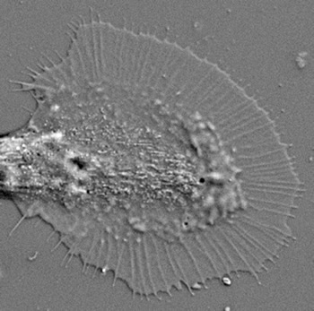
Aplysia growth cone in DIC optics. Diameter: 50 um. |
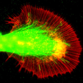
F-actin (red) and microtubule (green) labeling of the same growth cone. |
Regeneration following spinal cord injury (SCI)
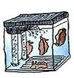
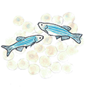
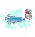
Regeneration of damaged axons in the central nervous system is a major challenge in adult mammals. We are interested in identifying new treatment approaches for SCI and conduct two projects in the area of axonal regeneration. In the first project, we are investigating the role of Nox-derived reactive oxygen species (ROS) in regeneration. In the second project, we use a visual motor response (VMR) assay and zebrafish larvae to screen a drug library. Our goal here is to identify new drugs that enhance axonal regeneration and functional recovery following SCI. Supported by a grant from the Indiana Spinal Cord and Brain Injury Research Fund.
 |
|
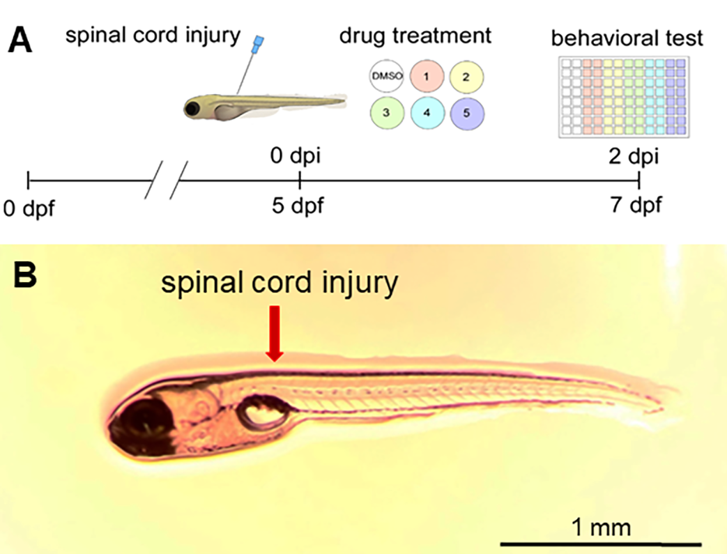
(A) Timeline of drug screen for zebrafish spinal cord injury (SCI). SCI is performed at 5 days postfertilization (dpf) followed by drug treatment for 2 d and VMR assay at 7 dpf to assess swimming behavior. (B) Location of SCI in zebrafish larva. |
Role of reactive oxygen species in regulating growth cone motility and development of retinotectal connections
Reactive oxygen species (ROS) are now recognized to have physiological signaling role besides causing oxidative damage. We have shown that upon lowering growth cone ROS levels in general, or by inhibiting specific sources such as NADPH oxidases and lipoxygenases, F-actin organization and dynamics are severely impaired (Munnamalai and Suter, 2009). These results suggest that localized ROS sources in the growth cone can regulate the growth cone actin cytoskeleton and related motility. We have further demonstrated that a NOX-2 type NADPH oxidase complex exists in Aplysia growth cones. NOX-2 exhibits an interesting bidirectional relationship with the actin cytoskeleton (Munnamalai et al., 2014). Follow-up studies in the developing zebrafish central nervous system revealed that Nox2 is involved in the formation of retinotectal connections (Weaver et al., 2018) and acts downstream of slit2/robo2 in retinal ganglion cell growth cones (Terzi et al., 2021). Ongoing studies address the function of Nox2 and ROS in the regulation of growth cone motility and guidance both in vitro and in vivo. Supported by NIH grant R01NS117701.
|
 |
|
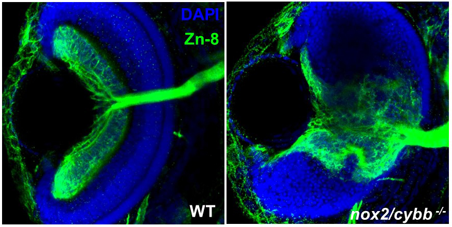
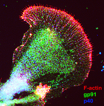
| Aplysia bag cell growth cone stained for F-actin (red), gp91phox (green), and p40phox (blue). Both gp91phox and p40phox exhibit partial co-localization with F-actin. From Munnamalai et al. 2014 | Eyes in WT and nox2-/- homozygous mutant zebrafish embryos at 72 hpf stained with DAPI (blue) and Zn-8 antibody (green), which labels retinal ganglion cells. The lack of functional Nox2 results in defects in layer formations in the retina as well as proper formation of the optic nerve. From Weaver et al. 2018. |
Neuronal mechanics
In collaboration with the lab of Dr. Arvind Raman, Mechanical Engineering, Purdue University, we have measured the retrograde traction force developed by Aplysia growth cones as they undergo adhesion-mediated growth cone advance. We have used two different approaches: force-calibrated microneedles and AFM. We have found that Aplysia growth cones can produce a wide range of retrograde traction forces (1-100 nN) as they advance in response to a local adhesion substrate. Our biophysical studies further revealed a linear relationship between substrate stiffness and force development suggesting a reinforcement mechanism (Athamneh et al., 2015). Ongoing studies address the nature of the seemingly constant substrate deformation observed during adhesion-mediated growth cone advance as well as mechanisms of mechanosensing.
|
 |
|
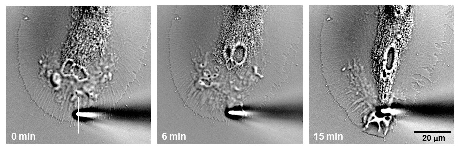
DIC images showing an apCAM-coated microneedle with 0.007 N/m stiff-ness interacting with a growth cone. The three images show the microneedle when first touching the growth cone (0 min), the C domain beginning reorien-tation toward the microneedle contact point (6 min; end of latency period), and C domain reaching the microneedle tip (15 min; end of traction period). From Athamneh et al., 2015. |